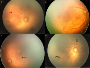
Retinopathy of Prematurity (ROP)
ROP is an eye disorder affecting premature infants, caused by abnormal retinal blood vessel growth. Early detection and treatment help prevent long-term vision loss
Retinopathy of Prematurity Incidence/Prevalence
Retinopathy of prematurity (ROP) is a potentially serious eye condition that affects premature infants, particularly those born before 31 weeks of gestation or with a low birth weight. It occurs when abnormal blood vessels grow in the retina, the light-sensitive layer at the back of the eye. This can lead to vision problems or even blindness if not treated. The prevalence of severe ROP (stages III and above) is lower, typically around 5% to 10% in high-risk populations. The incidence of ROP is estimated to range from 15% to over 50% among infants born before 28 weeks of gestation. Infants with very low birth weight (less than 1500 grams) have a higher incidence, often exceeding 50%.

Retinopathy of Prematurity Causes
- Prematurity
- The most significant risk factor; the earlier a baby is born (especially before 31 weeks of gestation), the higher the likelihood of developing ROP.
- Low Birth Weight
- Infants weighing less than 1500 grams (about 3.3 pounds) are at increased risk.
- Oxygen Therapy
- While oxygen is essential for premature infants, excessive or fluctuating oxygen levels can disrupt normal retinal blood vessel development, leading to ROP.
- Unstable Health Conditions
- Conditions such as respiratory distress syndrome, infections, and other complications in premature infants can contribute to the development of ROP.
- Genetic Factors
- Genetic predisposition may also play a role, although the exact genetic components are still being studied.
- Environmental Factors
- High levels of light exposure, fluctuations in temperature, and other environmental stresses in neonatal intensive care units (NICUs) may contribute to ROP risk.
- Anemia and Blood Transfusions
- Anemia is common in premature infants, and blood transfusions are sometimes necessary, which may increase the risk of ROP.
Retinopathy of Prematurity Diagnosis and Examination Process
The diagnosis and examination process for retinopathy of prematurity (ROP) is critical for early detection and intervention. Here’s an overview of the steps involved:
Diagnosis and Examination Process
- Eligibility for Screening:
- Infants born before 31 weeks of gestation or weighing less than 1500 grams are typically screened for ROP.
- Infants with additional risk factors may also be considered for screening.
- Timing of First Examination:
- The first eye exam usually occurs around 4 to 6 weeks after birth, depending on the infant’s gestational age and clinical status.
- Referral to an Eye Specialist:
- A pediatric ophthalmologist or retina specialist experienced in ROP performs the examination.
- Preparation for the Exam:
- The infant is usually positioned comfortably, often in an incubator.
- Eye drops are administered to dilate the pupils, allowing for better visibility of the retina.
- Examination Techniques:
- Indirect Ophthalmoscopy: The specialist uses a light source and a special lens to visualize the retina. This method allows for a detailed view of the retinal blood vessels and the overall condition of the retina.
- Retinal Imaging: In some cases, advanced imaging techniques like fundus photography or optical coherence tomography (OCT) may be used to assess the retina more thoroughly.
- Staging and Classification:
- The ophthalmologist evaluates the retina for signs of ROP and assigns a stage from 1 to 5 based on severity:
- Stage 1: Mild, with abnormal blood vessel growth.
- Stage 2: Moderate, with more pronounced changes.
- Stage 3: Severe, with more abnormal growth and potential complications.
- Stage 4: Partial retinal detachment.
- Stage 5: Total retinal detachment.
- The ophthalmologist evaluates the retina for signs of ROP and assigns a stage from 1 to 5 based on severity:
- Follow-Up:
- Infants diagnosed with ROP may require regular follow-up exams to monitor the progression or regression of the condition.
- The frequency of follow-up visits depends on the stage of ROP and the infant’s overall health.
- Documentation and Communication:
- The findings from the examination are documented, and results are communicated to the infant’s healthcare team, ensuring coordinated care.

Retinopathy of Prematurity Treatments
Treatment for retinopathy of prematurity (ROP) varies based on the severity of the condition. Here’s an overview of the common treatment options:
Treatment Options for ROP
- Monitoring:
- Observation: Mild cases of ROP may resolve on their own. Infants with Stage 1 or Stage 2 ROP are often closely monitored with regular eye exams to track any changes.
- Laser Therapy:
- Laser Photocoagulation: This is the most common treatment for more severe ROP (usually Stage 3 and above). A laser is used to destroy peripheral retinal tissue that is not adequately supplied with blood, which helps to stabilize the condition and prevent further abnormal blood vessel growth.
- Cryotherapy:
- Cryoablation: In some cases, freezing treatment may be used to destroy peripheral retina tissue. This method is less common now, as laser therapy has become the preferred approach.
- Anti-VEGF Therapy:
- Injections: Medications that inhibit vascular endothelial growth factor (VEGF) can be injected into the eye to help reduce abnormal blood vessel growth. This treatment is used for certain cases of severe ROP and may be combined with laser therapy.
- Surgical Intervention:
- Vitrectomy: For advanced cases (like Stage 4 or 5 ROP), where there is retinal detachment, surgical intervention may be necessary to repair the retina. This procedure involves removing the vitreous gel and may involve reattaching the retina.
- Long-Term Management:
- Vision Rehabilitation: Infants who experience vision impairment due to ROP may benefit from vision therapy and rehabilitation services to support their development and adaptation.
Consult an Eye Expert for Your Baby Today

Doctors and Specialists
Our Medical Team
Our team of highly skilled and experienced ophthalmologists and optometrists is committed to delivering personalized, comprehensive eye care

Phaco Surgeon & Vitreo-Retinal Surgeon
Dr. Mohamed Azzam

Vitreo-Retinal & Phaco Surgeon
Dr. Amogh Dileep Asgaonkar

Phaco Surgeon and Pediatric Ophthalmology
Dr. Arjun Malla Bhari

Testimonials
Read inspiring stories from patients who have experienced clearer vision and compassionate care with EyeCare, reflecting the trust and results we strive for every day
Frequently Asked Questions
A. ROP is classified into five stages, ranging from mild (Stage 1) to severe (Stage 5), with increasing severity indicating a greater risk of vision loss.
A. Yes, mild cases (Stages 1 and 2) often resolve without treatment. Close monitoring is essential to ensure that any progression is addressed promptly.
A. Parents should discuss any concerns about their infant’s vision or eye health with their pediatrician or a pediatric ophthalmologist, especially if the infant is at risk.
A. Early signs are often not visible to parents, as ROP typically doesn’t show symptoms until later stages. However, signs of vision issues, such as unusual eye movements or difficulties with focus, may be observed as the infant grows.
A. ROP primarily affects premature infants, but in rare cases, full-term infants with certain health conditions may also develop similar retinal issues.
A. Depending on the severity, ROP can lead to various vision problems, impacting visual development. Early intervention and rehabilitation can help support visual skills.

